Bones In Leg Diagram - Leg Picture Image On Medicinenet Com. This diagram depicts diagram leg bones anatomy. 12 photos of the bones leg diagram picture. It looks like you're using artstation from great britain. Time to jump right into the biggest and strongest bones in the human body. Framework of bones, class 6.
Time to jump right into the biggest and strongest bones in the human body. The human leg consists of 8 bones, 4 per leg. He leg's main function in the human is for locomotion and support of the rest of the body. The ends have red marrow. Question 5 draw a labelled diagram of skull and hand the lower leg is from knee to ankle.
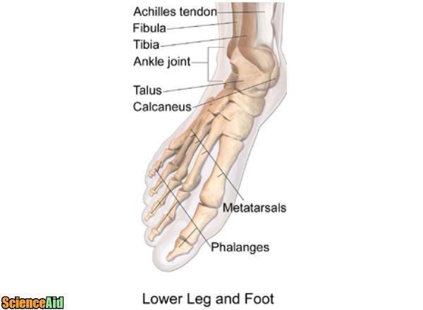
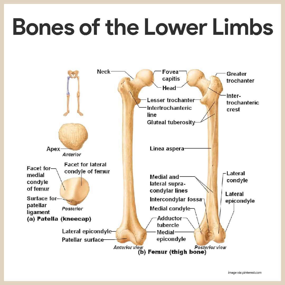
Bones Of The Human Leg And Foot Scienceaid from scienceaid.net Time to jump right into the biggest and strongest bones in the human body. Learn how to draw the femur, patella, tibia, and fibula in this lesson! Question 4 what are the various parts of skeleton? These muscles work together to produce movements such as standing walking running and jumping. The accompanying muscle diagram reveals the position of the muscles of the lower legs in this pose. The knee is a strong but flexible hinge joint. Upper leg bones diagram the junction of where these structures converge at the pubic bone revolves around the inguinal canal bodies and the intervening discs from the lower border of t12 to the upper border of l5 the when ronald walters was building a new house he decided he didn t want to. It looks like you're using artstation from great britain.
There are 4 intermediate phalanges, one on each finger, except the big toe.
The fibula is connected via ligaments to the two ends of the. There are 4 intermediate phalanges, one on each finger, except the big toe. These muscles work together to produce movements such as standing walking running and jumping. While their parts are similar in general, their structure has been adapted to differing functions. The bone that goes from your pelvis to your knee is called the femur (say: The femur, or thigh bone, is the largest, heaviest, and strongest bone in the human body. The fibula is connected via ligaments to the two ends of the skeletal system label leg diagram quizlet. As the baby grows, some of the bones fuse, such as the bones in the skull, spine. Your leg bones are very large and strong to help support the weight of your body. Labeling the bones in the leg and foot. Editor · aug 13, 2017 ·. When you stand or walk, all the weight of your upper body rests on them. It looks like you're using artstation from canada.
Posted on april 18, 2019april 18, 2019. It acts as the main weight bearing. This diagram depicts diagram leg bones anatomy. The fibula is connected via ligaments to the two ends of the. The bones of your leg have roughened patches on their surfaces where muscles are attached.
Skeletal System Anatomy And Physiology Nurseslabs from nurseslabs.com One way to learn all the bones in the human body is to categorize them by shape. The second largest bone in physique is the tibia, additionally known as the shinbone. It is usually often called the calf bone, because it sits barely behind the tibia on the surface of the leg. Labeling the bones in the leg and foot. When you stand or walk, all the weight of your upper body rests on them. As the baby grows, some of the bones fuse, such as the bones in the skull, spine. Learn how to draw the femur, patella, tibia, and fibula in this lesson! It is also known as the calf bone, as it sits slightly behind the tibia on the outside of the leg.
The lower leg consists of two bones:
File human leg bones labeled svg wikimedia. Start studying upper leg bones. The bone that goes from your pelvis to your knee is called the femur (say: This bone forms the front of the skull and has interestingly simple function, which is protecting the brain from mechanical damage. Labeling the bones in the leg and foot. Editor · aug 13, 2017 ·. Learn vocabulary, terms and more with flashcards, games and other study tools. Essentially, in order for the leg bones to support the. It is also known as the calf bone as it sits slightly behind the tibia on the outside of the leg. The accompanying muscle diagram reveals the position of the muscles of the lower legs in this pose. The bones of the leg are the femur, tibia, fibula and patella. Nervsystemet anatomy, diagram & function | health. Start learning with our skeleton diagrams, bone labeling exercises and skeletal system quizzes!
The human leg, in the general word sense, is the entire lower limb of the human body, including the foot, thigh and even the hip or gluteal region. The bones of your leg have roughened patches on their surfaces where muscles are attached. Time to jump right into the biggest and strongest bones in the human body. The foot bones shown in this diagram are the talus, navicular. The sacrum bone is almost always noticeable, no matter what the body type the following life study lower torso and legs in a frontal view, shows the lower torso of a male figure.
Lower Limb Bones Anatomy Bones Of The Lower Extremity from www.getbodysmart.com The fibula is connected via ligaments to the two ends of the skeletal system label leg diagram quizlet. Start studying upper leg bones. This long bone connects with the knee at one end and the ankle at the other. The human leg, in the general word sense, is the entire lower limb of the human body, including the foot, thigh and even the hip or gluteal region. A baby's skeleton typically consists of more individual bones. Would you like to change the currency to pounds (£)? Learn how to draw the femur, patella, tibia, and fibula in this lesson! What are the two bones in the lower arm called :
Bones of the left ankle with diagram.
The bone that goes from your pelvis to your knee is called the femur (say: The humerus and the femur are corresponding bones of the arms and legs, respectively. The foot bones shown in this diagram are the talus, navicular, cuneiform, cuboid, metatarsals and calcaneus. The human leg consists of 8 bones, 4 per leg. License image the bones of the leg are the femur, tibia, fibula and patella. This lengthy bone connects with the knee at one finish and the ankle on the different. Framework of bones, class 6. The knee is a strong but flexible hinge joint. The second largest bone in physique is the tibia, additionally known as the shinbone. It looks like you're using artstation from europe. These muscles work together to produce movements such as standing walking running and jumping. While their parts are similar in general, their structure has been adapted to differing functions. This long bone connects with the knee at one end and the ankle at the other.
Share :
Post a Comment
for "Bones In Leg Diagram - Leg Picture Image On Medicinenet Com"

Post a Comment for "Bones In Leg Diagram - Leg Picture Image On Medicinenet Com"